Part Two: The Seat of Personality
This chapter marks a transition in this book. The transition is from everyday English language and concepts, to the specialized language and concepts of neuroscience.
The Introduction said that this book serves as a vehicle for translating and matching the Enneagram and neuroscience. There, I said that the Enneagram authors tend to speak in terms of esoteric concepts like "growth dialectic", "return to essence", and "spiritual work." But as you've seen, this book has pretty much steered clear of that sort of language in describing the Enneagram.
In describing the Enneagram, this book has introduced the fairly straightforward concepts of fear, triads, development, security, and wings. These concepts were then analyzed for the purpose of squeezing out of the Enneagram four fundamental notions: optimism, pessimism, aware fear, and unaware fear.
These latter notions are the lynchpin of this book. While Part One shows how these notions fall out of the Enneagram, Part Two shows how they relate to neuroscience.
But to understand how neuroscience treats these notions, we need to learn a whole new language. At least we need to learn some key concepts in this new language. These key concepts include the triune brain, brain asymmetry, prefrontal cortex, amygdala, DNA, genes, glucose metabolism, neurotransmission, brain plasticity, PET, fMRI, and EEG. If you are unfamiliar with neuroscience, much of this list may seem like Greek to you.[1]
The remaining chapters of this Part will rely on these concepts. If you are a neuroscientist and these concepts are familiar to you, you may want to skip this chapter. Alternatively, you may want to read it to check whether the foundation of this book's hypothesis is solid. But for everybody else, you need to read this chapter to understand the rest of this book.
Chapter 1 introduced the Enneagram basics; this chapter offers a primer on the brain. Just as Chapter 1 may have seemed less interesting than other chapters, the same may hold true for this chapter. But as our mothers used to say to us: "I know it tastes bad, but it's good for you."
The brain is one of the organs in our bodies. Other bodily organs include the heart, the liver, and the lungs. As with these other organs, the brain has a specific function. Where the heart serves to pump blood, the liver to detoxify the body, and the lungs to process the air we breathe, the brain serves as the master controller for the entire body.
Our brains constantly monitor the functioning of every corner of our bodies. They also play a principal role in driving the activity going on in our bodies. This includes conscious activity. For example, the voluntary typing action of my fingers on this keyboard is being driven by my brain. But also, the brain controls unconscious activity in the body. For example, heat rate, digestion, metabolism, immune functioning, and breathing are all, to one extent or another, under the master control of the brain.
This book focuses on only a few aspects of the brain's functioning. Specifically, this book looks closely at the brain's control of mood and fear.
The brain is a three-pound organ resting inside our skulls. But this organ is not an amorphous three-pound blob. Instead, it consists of reasonably discrete parts connected together, kind of like a toy truck constructed of Lego blocks.
Since the brain is a three-dimensional object, discussions about its various parts can become complex. To simplify things, it helps to look at these parts from three different viewing perspectives: layers, hemispheres, and regions.
The first viewing perspective concerns the layers of the brain. Like an onion that is composed of various layers, so the brain is also made up of layers. The multiple layers of the brain are grouped into three principal structural layers: base, limbic system, and cerebral cortex.
The base of the brain is situated on top of our spinal column. This is the deepest of the layers. On top of and enveloping this base is the so-called "limbic" system. This is the middle layer. Finally, the outer layer, the cerebral cortex, covers the limbic system.
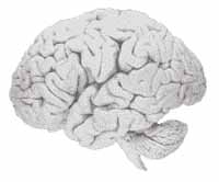
Common pictures of the brain resemble a gray lump of cottage cheese. This "cottage cheese" is the cerebral cortex. Although it may seem from these pictures that the brain is entirely made up of this cottage cheese, this not the case. The cerebral cortex is a relatively thin layer covering the rest of the brain like a skull cap. Moreover, in a living brain, this skull cap is not gray, but rather reddish in color. This is because living brains are infused with blood.
This three-layer system is known in the literature as the "triune" brain. "Triune" just means "being three in one." Before the recent invention of advanced brain technologies that allow detailed study of living brains, it was assumed that this triune brain did not merely comprise discrete structural layers. It was thought that these discrete structural layers also corresponded to discrete functions. In other words, it was thought that because these layers looked quite different from each other, they also did quite different things.
This early line of thinking held that the base of the brain is "reptilian", the limbic system "mammalian", and the cerebral cortex "human". The notion was that reptiles possess only the base layer in their brains; mammals have both the base layer and the limbic system, but not the cerebral cortex; and humans are uniquely blessed with all three layers.
This earlier line of thinking was based on studying the dead brains of dissected animals. Because dead brains don't function, all that was available for studying at that time was structure. And the structure hinted strongly at a "triune" brain with distinct function corresponding to the apparently sharp differences among the various animal species.
Specifically, "tradition" has it that the base of the brain corresponds to instinct; the limbic system to feeling; and the cerebral cortex to thinking. This is why, if you perform an Internet search for "triune brain thinking feeling instinct," you will be presented with many pages describing this triune hypothesis.
The problem, however, is that this hypothesis has proven incorrect. The new brain technology of the last few decades has swept the earlier "triune brain" hypothesis into the dustbin of discarded scientific notions. Because this recent technology is able to study living brains at a cellular level, scientists now have the ability to understand brain functioning.
What this recent research shows is that the triune brain is a fiction from the point of view of gross, simplistic function. Sure, the three layers are structurally different. But functionally, the layers work together to perform thinking, feeling, and instinct. This is especially clear for the mental activity of feeling.
Joseph LeDoux of New York University is among the world's leading researchers on the neuroscientific basis of emotion. Mr. LeDoux has authored a pair of recent books, The Emotional Brain: The Mysterious Underpinnings of Emotional Life and Synaptic Self: How Our Brains Become Who We Are. These books summarize his research and synthesize the field.
In his books, Mr. LeDoux makes clear that the systems in our brains that process and generate emotions and feelings span both the cerebral cortex and the limbic system. Thus "feeling" cannot be said to be a "limbic system" function.[2] Indeed, the conscious component of feeling — i.e. the state of being aware of our feelings — is likely found principally, and perhaps solely, in the cerebral cortex. So the triune brain is not particularly useful as a functional concept. However, as a structural description of the brain, it remains an accurate notion.
The other reason the triune brain concept is not useful concerns its "reptilian, mammalian, human" triad. As noted, this concept assumes that reptiles do not have limbic systems or cerebral cortices, and that non-human mammals lack cerebral cortices. Recent research has demonstrated these assumptions to be incorrect. All vertebrates have all three layers. But each layer is developed to different degrees for each animal. Humans possess by far the most developed cerebral cortex. Although the cerebral cortex of the snake is largely undeveloped, it does exist. This existence was confirmed through the microscopic study of living snake brains. [3]
The fundamental point here about the brain layers is that brain function is much more complex than the relatively simple brain structure. Indeed, studying function reveals the brains of humans to be much closer to the brains of other animals than was previously thought in many circles.
The second viewing perspective concerns the two halves of the brain. Let's say this is a horror movie, and the villain runs a skill-saw down the middle of our forehead. The cut cleaves our head in half, with one ear attached to each half. The cleave would reveal two "mirror-image" halves of the brain. It's like a walnut split cleanly in half along the seam of the shell.
These two halves are referred to as the "left hemisphere" of the brain, and the "right hemisphere" of the brain. More simply, the terms "left brain" and "right brain" are used. In the past, these phrases were often meant to refer only to the cerebral cortex. In other words, "left brain" referred to the left half of the cerebral cortex. The limbic system was considered to be more unified in function, even though it is also structured as two halves.
Recent studies have emerged showing that the limbic system does seem to exhibit subtly different function for the left and right sides. Different function for each half is called "hemispheric asymmetry". "Hemisphere" literally means "half of a sphere;" "Asymmetry" means "not identical". Beyond the cerebral cortex and the limbic system, asymmetry has also been observed in the base of the brain.
Not only are the two halves of the brain different in function, they are also not structurally identical. Small differences in shape and size have been observed between the left and right sides of the brain. Indeed, these two halves develop at different rates during fetal and infant development.
The third and final viewing perspective concerns the regions of the brain. Each layer of the brain is composed of different, fairly discrete regions. It's like a map of the world. All of the landmass can be considered one big collection of land. But at a next level of analysis, the landmass can be divided into different countries, each defined by a border. Deeper still, most countries can be divided into states or provinces. These states can be divided into counties; the counties into towns and cities; the towns and cities into streets; the streets into properties; and so on.
This top-down methodology is similar to how the regions of the brain are classified. As with a map of the world, the regions and sub-regions and sub-sub-regions are far too many to comprehend in one sitting. However, for the purposes of this book, we will be looking at only two regions of the brain: the "prefrontal cortex" and the "amygdala".
The prefrontal cortex is a region of the cerebral cortex. This region is located in the front of our foreheads. Actually the region is composed of a left half and a right half. The left prefrontal cortex sits just above our left eye; the right prefrontal cortex just above our right eye.
The amygdala is a region of the limbic system. As with the prefrontal cortex, the amygdala consists of a left half and a right half. Each half resembles an almond. Indeed, "amygdala" is the Greek word for almond. These two almonds lie inside our temples. The right amygdala lies about one inch inside our right temple; the left amygdala about one inch inside our left temple.
The reason this book focuses on the prefrontal cortex and the amygdala is that current research shows these two regions playing the biggest roles in processing and generating the mental states of optimism and pessimism (prefrontal cortex), and aware and unaware fear (amygdala and prefrontal cortex).
Movies tend to feature many different actors playing many different roles. But in most movies, there are only a couple of major stars who play the biggest roles in the movie. They have the most lines, and the plot centers around their characters. Still, many other lesser characters are required to fill out the whole movie.
In a similar way, the prefrontal cortex and the amygdala are not the whole movie when it comes to mood and fear. Many other regions in the brain collaborate with these two "starring" regions to process and generate these mental states. But discussing all of these "supporting" regions would increase the complexity of the discussion without improving it.
So this book trains its focus on the stars of the show: the prefrontal cortex and the amygdala. Chapter 5 is devoted to the prefrontal cortex; Chapter 6 to the amygdala.
The preceding section says that the prefrontal cortex and the amygdala play the lead roles in "processing and generating" the mental states of optimism, pessimism, aware fear, and unaware fear. But it doesn't say how these regions do this processing and generating. To understand this, we need to understand neurons. But to understand neurons, we first need to understand cells.
A neuron is a particular kind of cell. The human body is made up of billions of tiny cells. Every cell in our body is a discrete structure. The outer boundary of the cell is defined by a wall. The cell wall encloses the entire cell.
Communication between cells is accomplished by the passage of particular molecules in and out of the cell wall. The cell wall is like the immigration authorities at the U.S. border. If the system works as intended, nobody comes into the U.S. without showing proper papers. Some people are denied entry. Similarly, the cell wall watches closely to ensure that only appropriate molecules pass into the cell.
Every cell also contains a central command center known as the nucleus. The nucleus is a discrete, enclosed region floating within the bounds of the cell wall. Inside the nucleus of every cell in our bodies is a twisted pair of long molecules known as "DNA".
DNA ("deoxyribonucleic acid") is like a master set of recipe books for the entire body. That is, DNA contains all the instructions necessary for generating and operating our entire body. This is an enormous collection of information.
Recently, the Human Genome Project announced that it had mapped the human genome. What that means is that if human DNA is like a set of recipe books, the project identified the sentences and words that make up those books. Previously, the words and sentences were not apparent. DNA seemed to be just a virtually endless stream of characters. But the project figured out how to properly break up this stream into sentences and words. In the language of computer science and compilers, this is called "syntax parsing."
The next step is to figure out what the sentences and words mean. Scientists all over the world are racing to decode the genome. Every month it seems we read about the discovery of the "gene for trait X". What this means is that the "words and sentences" in DNA that generate trait X may have been isolated. In computer science, this step is known as "semantic analysis."
Finding the words and sentences that correspond to a particular trait is no easy feat. At the current state of technology, this is the classic "needle in the haystack" problem. The few words and sentences that generate the trait are the needle amidst the haystack of the billions of words and sentences that make up DNA.
Saying that certain words in DNA "generate" a trait may sound strange. It's like saying that a particular recipe for a cake makes the cake. But that's not true. Letters on a page can't mix the flour, butter, and milk. For that, a cook is needed. The cook reads the recipe, and follows the instructions to make the cake.
In the cellular world, the cooks are the enzymes. Enzymes "read" certain words within DNA, and go about the work of performing the instructions indicated by those words. This is why enzymes are so important. No cooks, no meals; no enzymes, no life.
Now the steps from enzymes reading "words" within DNA, to the manifestation of gross traits traceable to DNA (such as eye color), are a great many. It is well beyond the scope of this book to describe these lengthy biochemical paths. Just understand that there are such paths. So DNA, as the mouth of a great river that flows toward generating much of who and what we are, is an interesting subject to ponder.
When we say that a certain trait, such as eye color, is "genetic," we mean that this trait is encoded directly in DNA. The opposite of genetic in this regard is the notion of "environmental". Environmental traits are those that are traced to experiences in our lives, and not directly to DNA.
The difference between genetic traits and environmental traits is crucial because the former are very difficult if not impossible to change, whereas for the latter, change is at least theoretically possible. Within the realm of personality theory, this distinction is crucial. What about ourselves can we change? And what, for better or worse, are we stuck with?
Now although every corner of our body is made up of cells, different corners often contain different kinds of cells. These different kinds of cells perform different functions. These different functions often demand very different shapes.
Muscle cells tend to be very long, thin, and tubular. The function of a muscle cell is to allow the muscle to contract and release. Muscle contraction is accomplished through the "bunching up" of the cells in that muscle. In contrast, liver cells are shaped like beads and arranged in strings of these "beads". Liver cells serve to process toxins in the body.
Neurons are the cells that make up the brain. Neurons have a main body in which the nucleus (containing DNA), as well as other items, is stored. From this main body, multiple thin projections reach out to other neurons.
There are two main kinds of these projections: axons and dendrites. Each neuron typically has only one axon, although the end of the axon can branch out to connect with many other neurons. The function of the axon is to send signals to other neurons. The axon is the "output" pathway for the neuron.
Dendrites are the "input" pathways for the neuron. It is via its dendrites that a neuron receives signals from other neurons. So signals run from the dendrites, through the main body of the neuron, and on out along the axon. Some neurons have thousands of dendrites.
These "signals" are electrical currents. This electricity in our brains is the same kind of electricity that runs through our houses. Electricity is just the flow of electrons. But the difference is that the electrical activity in our brains is a tiny fraction of the level required to run our toasters.
So one part of the story of how our brains operate concerns electrical currents running through our brains conveying messages. But there is more to this story. Unlike the wire of our toaster that is a single cord, "wires" in our brain consist of multiple discrete neurons. And these neurons do not touch each other. If they don't touch each other, how does electricity flow from one neuron to the next?
The problem of "neurotransmission" now shifts from a matter of electrical engineering, to one of chemical engineering. A "sending" neuron communicates with a "receiving" neuron by way of the axon of the sending neuron connecting to a dendrite of the receiving neuron. But the sending axon and the receiving dendrite do not physically touch. Instead, there is a tiny gap between them. This gap is known as the "synapse".
In the synapse, lots of interesting chemical things happen. Essentially, certain chemicals, known as "neurotransmitters", travel from the axon to the dendrite. That is, the electrical current running along the axon causes the axon to start a chemical process resulting in the transmission of certain neurotransmitters to the dendrite. Receiving these neurotransmitters causes the dendrite to start a process that results in an electrical current being generated and sent along the dendrite toward the main body of the receiving neuron.
Neurotransmission within the synapse is interesting because this is the target of many different kinds of brain medications. For example, certain kinds of anti-depressants, like Prozac, prevent the removal of serotonin from the synaptic space. Serotonin is one kind of neurotransmitter that is used for communication between neurons. Depression can act to deplete certain areas of the brain of serotonin, and Prozac acts to nudge serotonin to stay in the brain for use in neurotransmission.
Of course, this begs the question: What caused the depression that led to the depletion of serotonin? Presently, this is an open question in medicine. If the hypothesis of this book is correct — that the Enneagram is reflected in the workings of the brain — medical mysteries like this one may soon be solved.[4]
The previous section described an electrical charge running through a neuron to result in a neurochemical exchange with a receiving neuron. Energy is required to generate this electrical charge, as well as to conduct this neurochemical exchange. It's the same principle as the electricity that runs our toasters. That electricity has to originate from some energy source. In our electrical grid, these sources are the electrical power plants. Those power plants get their energy from the processing of certain kinds of fuel, including petrochemicals, radioactive materials, sunlight, the wind, water flow, and many other kinds of fuel.
In neurons, the power plant is contained in the cell body, and the fuel "burned" in the power plant is glucose. Glucose, which is a form of sugar, enters our bodies via the food we eat. From our small intestines, glucose from our food enters our bloodstream. Blood travels to our brain and feeds glucose to our brain. In other words, our brains have a serious "sweet tooth".
The power plant of the neuron "burns" glucose to generate the energy required for neurotransmission. This burning is referred to as "metabolism". "Glucose metabolism" in the brain refers to the process by which glucose is chemically converted into a form of energy usable by the neuron to function.
It is important to note that blood entering a certain part of the brain is an indication that neurotransmission is going on there. The brain doesn't hoard blood or glucose, or the intermediate by-products of glucose metabolism. Instead, the brain employs a "just-in-time" inventory system for glucose. Once glucose enters the neuron, it goes straight into the production of energy resulting in neurotransmission.[5]
The previous section concludes the discussion about what the brain consists of and how it works. Now the discussion shifts to how the working of the brain is measured. How can we tell what is going on in living brains?
Before the last couple of decades, the answer to this question was: "With difficulty." That is, methods available to those early researchers did not offer a full and clear view into the living brains of humans. Constrained by these limitations, neuroscience as a field crept along, with advances measured on the order of decades or even centuries. But today, with the current technology, advances in brain science are racing forward almost as fast as you are reading this.
In the old days, one way to try to figure out how the brain worked was to study people with brain damage. One of the most famous cases was that of a man named Phineas Gage. A mild-mannered delivery-man, Mr. Gage one day suffered an accident whereby a metal rod pierced the left frontal side of his skull. He survived the accident, but with the hole in his head, his personality changed radically. He became coarse and aggressive. Brain researchers had a field day with this fellow.
The limitation with this method of research was that the researchers could not be sure of exactly which regions in Mr. Gage's brain were damaged. Researchers had to wait until he was dead at which time an autopsy was performed and revealed the extent of the damage. But by that point, no further research could be conducted since the man was dead. What this means is that brain research involving the study of brain-damaged patients like Phineas Gage was largely a case of "shooting in the dark". Moreover, using this method of research, advances in knowledge proceeded on the order of centuries.
A second older method of brain research involved the use of animals. With animals, researchers could damage the brains of the animals in specific ways to test the effect of the damage. This is, in fact, how the electrical aspect of neurotransmission was discovered. A researcher attached an electrical probe to the brain of a frog and noticed that the frog's leg twitched. Thus it was first discovered that electricity is one means by which brains function.
The limitation with this method of research was and is that animals are not humans. Before living human brains could be studied in detail, studying animal brains and trying to tie the results of that research to the human brain was a reasonably speculative enterprise. Researchers suspected that some aspects of animal brains worked reasonably the same as certain aspects of human brains. Other aspects were different. But how to know which was which?
Still another older method of human brain research is known as EEG (electroencephalogram). Given that neurotransmission involves the flow of electricity, this electrical activity is subject to measurement. This is precisely what EEG does. EEG involves the placement of electrodes (small metallic discs) at various locations on the scalp of the patient. These electrodes pick up on the electrical activity occurring in the brain regions just under the skull.
The limitation of this method of research was and is that EEG had and still has difficulty measuring electrical activity occurring deep within the brain.[6] This includes activity in the amygdala. As we'll see, activity in the amygdala is critical to the neurological understanding of human personality. So armed only with EEG, a researcher could not get a full picture of human personality.
The last quarter of the twentieth century witnessed breakthroughs in the study of living human brains. Two technologies were invented allowing unprecedented visibility into this mysterious, complex, and beautiful domain. These two technologies are known as PET and fMRI.[7]
PET, "Positron Emission Topography", was invented chiefly by Michael Phelps in 1973. That doesn't mean that by 1974, brain researchers were using PET, or that PET was sufficiently developed at that point to provide terribly useful information. Note that the Internet was invented by an arm of the U.S. Military in the 1960s. But it wasn't until the mid-1990s, with the emergence of the Netscape Internet Browser, that the Internet began taking off in terms of popular use. Similarly, PET did not take off among brain researchers until the mid-1990s. So the brain studies you will be reading about in this Part that rely on PET all come from the last few years.
fMRI, "Functional Magnetic Resonance Imaging," is an even more recent invention. fMRI emerged in 1990 out of AT&T Bell Laboratories. fMRI represented an advancement over an earlier, established technology, MRI. By 1990, MRI was fairly ubiquitous within big hospitals and research laboratories. So the adoption of fMRI was relatively rapid. Just as the adoption of PET was starting to pick up steam in the mid-1990s, so did the adoption of fMRI. So the brain studies you will be reading about in this Part that rely on fMRI also come from the last few years.
Both PET and fMRI allow the study of living human brains, and both use the fact of glucose metabolism to perform that study. Recall that glucose metabolism is an indicator of brain activity. In this way, PET, fMRI and EEG all ultimately study the same thing — brain activity — but they get there via different roads. EEG gets there via electrical activity; PET and fMRI via glucose metabolism.
Although PET and fMRI both study glucose metabolism, the way they do so is quite different. These differences account for different relative strengths and weakness of the two technologies. So some researchers — in particular well-funded labs — employ both technologies in the same study. They do this to get "the best of both worlds."
PET works via the use of radioactive substances. Radiation involves the emission of subatomic particles (electrons and positrons) from a molecule. Patients ingest a small, purportedly non-harmful amount of the radioactive substance, via injection or orally. Then the blood stream carries this substance to the brain, where the radioactive emissions are recorded by PET.
As noted, PET can study glucose metabolism. This is enabled by using a radioactive substance that interacts with glucose. The substance can be thought of as "piggybacking" on glucose, so when glucose is metabolized in the brain, the substance is there to let PET know about it.
PET is more flexible than fMRI in that it is not limited to the study of glucose metabolism. Different radioactive substances can be chosen to interact with different aspects of brain activity. For example, instead of attaching to glucose, a substance can be chosen that attaches to a neurotransmitter like serotonin. In that case, PET can be used to watch how the brain is making use of serotonin. As we saw earlier, serotonin is the target of one class of anti-depressants. But in this book, the studies you will be learning about all concern glucose metabolism.
fMRI works differently. fMRI, unlike PET, is "non-invasive". This means no liquid or solid substance is transferred into the patient to enable study. Instead, fMRI works through the use of a magnetic field. That is, with fMRI, the heads of patients are placed in a magnetic field. This field causes certain molecules in the brain to "line up" in such a way that fMRI can record the activity. Specifically, in brain studies, fMRI records the process of "blood oxygenation." The process of glucose metabolism — which as we've seen results directly in electrical and neurochemical brain activity — occurs via oxygen interacting with glucose to enable the release of energy. This is analogous to how wood requires oxygen to burn and thereby release energy in the form of heat. fMRI can be thought of as watching closely for the "smoke signals" of glucose metabolism.
Because of the differences in core technology between PET and fMRI, the two offer different strengths and weaknesses. Putting aside the ability of PET to measure processes other than glucose metabolism, one advantage of PET over fMRI is that PET allows measurement of blood volume, not merely blood flow. Blood flow is a transient measure of how much blood is moving through a particular area at a particular time. Blood volume indicates how much total blood has flowed through that region over a period of time. So PET makes it easier to get an answer to the question: How active has this particular region been?
On the other hand, fMRI has its advantages over PET. These include superior temporal resolution. You can think of this as shutter speed in a camera. How many pictures can you take in a short span of time? fMRI can take more pictures than PET. Also, fMRI allows better precision in identifying the region being studied. Think of this as the "focus" feature of the camera.
As noted, these relative strengths and weaknesses lead some researchers to use both technologies in certain studies. Those researchers are the lucky ones because PET and fMRI are very expensive technologies. The cost of full systems runs into the multi-millions of dollars.
A significant drawback of both PET and fMRI is that the patient needs to remain absolutely still while the recording is taking place. This makes it difficult to conduct "natural" experiments in "natural" settings. This is particularly so with experiments which seek to record emotional responses. In responding emotionally to a stimulus, we all tend to make certain bodily motions as part of that reaction. But these motions can interfere with the operation of PET and fMRI. So researchers do the best they can to get the patient comfortable and feeling reasonably natural, even though the patient is strapped to a gurney and placed in a space age contraption.
Even with this drawback, PET and fMRI represent dramatic improvements over the earlier methods of brain study — study of brain-damaged patients, animal research, and EEG. But these new technologies did not render these older methods obsolete. In fact, the opposite is true. PET and fMRI have rendered these earlier methods much more useful than before.
For example, consider the study of brain-damaged patients. In the old days, the problem with this method of research was that the researcher could not know the precise extent or location of the damage. But now, with PET and fMRI, the researcher knows a tremendous amount about the specific damage to any particular patient. With this knowledge, the information now coming out of the study of brain-damaged patients is tremendously useful.
Similarly, PET and fMRI have allowed researchers to understand the line separating animal brains from human brains. On one side of the line is similarity; on the other difference. Knowledge of where this line is has made the animal research much more useful in the understanding of the human brain. Richard Davidson warns researchers to mind this human-animal line that is now clear to all:
On the one hand, the rodent data have been essential in establishing some of the fundamental facts of subcortical circuitry ... . The convergence of aspects of the rodent work with studies of humans has been crucial for progress in this field. However, we now know that many of the anatomical details of crucial components of this circuitry are different in rodents and primates. The organization and connectivity of amygdala nuclei are different ... , [and] the anatomy of the prefrontal cortex is fundamentally different ... . These differences in anatomy imply differences in the nature, function, and complexity of emotions across species.[8]
Although PET and fMRI represent dramatic improvements over and to the older methods of brain research, these improvements have come at significant financial cost. The older technologies were available to "hacks" like myself. In this book, I am proposing a connection between the Enneagram and neuroscience. Why am I offering merely a hypothesis, instead of doing the research myself and offering the results of that research?
The answer is that I don't have a PET or fMRI system out back in my garage. I doubt that hardly any individual does. To use these systems to conduct the research needed for establishing a connection between the Enneagram and neuroscience, or for any other purpose for that matter, one needs to approach the institutions that own these systems.
Imagine a future in which a device the size of a football or motorcycle helmet can be fitted on the head of the patient. The patient can run around, flail his arms, do somersaults, or whatever. And the helmet provides all the information about the working of the living brain that PET and fMRI provide today. Since we're in fantasyland, imagine that this helmet runs for $79.99 at Wal-Mart. What would happen? One thing would be that we'd witness breathtaking "Stupid People Tricks" with the device. But, in addition, just imagine the advances in human knowledge. Maybe we'll see it in our lifetime.
But for now, back in present reality, we have to knock on the doors of the houses in which PET and fMRI live. "Can PET or fMRI come out to play?"
For the most part, PET and fMRI machines are owned by hospitals, and by universities in close association with hospitals. Why are hospitals the common element? The answer begins with a familiar injunction: follow the money.
PET and fMRI systems are very expensive. So the source of the money to pay for these systems becomes a significant consideration. In the vast majority of cases, the high cost of these systems means that an entity needs to earn income from these systems to pay for those costs. But how would these systems earn income? The answer is: health care costs.
CTI Molecular Imaging, Inc., of Knoxville, Tennessee, is a leading producer of PET systems. As CTI explains:
One of the principal reasons for growth in the PET market is the increasing number of PET applications being approved for reimbursement by third-party payors, such as Medicare, Medicaid, private insurers and HMOs. Reimbursement is critical to increasing the adoption rate of PET by clinicians.[9]
This book concerns only the use of PET and fMRI in brain studies. However, these systems are actually more often used in the treatment of diseases outside of the brain, including heart disease and cancer. Anyone reading the news knows there is a virtual epidemic of these sorts of diseases in the U.S. today.
The studies you will be reading about in the next two chapters were conducted primarily by professors from major universities that are associated with a hospital. Within these universities, these professors tend to work in the departments of psychology, neurobiology, or medicine.
This list of university departments is interesting in itself. Prior to PET and fMRI, our collective understanding of the human brain was relatively dim. Accordingly, significant overlap among the fields of psychology, biology, and medicine were not so obvious and were certainly not universally accepted. But now, these three types of professors bump into each other regularly in the PET and fMRI labs at their schools. Starting from different origins, they all meet at the same destination: the workings of living brains. It a fundamental purpose of this book to get the Enneagram experts invited to the party.
The other interesting dynamic going on here concerns my role. I am neither a professor nor even a student of psychology, neurobiology, or medicine, nor am I an Enneagram expert. So what the heck am I doing writing this book?
The answer has to do with the Internet. Prior to the Internet, it would have been quite difficult for me to write this book. One reason is that it would have been difficult to get hold of the neuroscience papers you will be reading about in the next two chapters. Before the Internet, these papers were accessible only from within the universities and the hospitals. I would have had to obtain permission from one of those institutions to use their information systems.
The problem is even deeper than that. If these papers had not been on the Internet, I wouldn't have even known what to look for. That is, it probably would not have occurred to me to even seek permission at universities or hospitals to conduct research. The reason is that the ideas in this book evolved over the course of reading a number of the studies. It was only after reading a sufficient number of them that the proverbial "light bulb" went off in my head, and spurred me to write this book.
Let's assume for a moment that the hypothesis of this book is correct and that it will prove useful to humanity. Certainly, that prospect has excited me enough to fund my motivation for writing this book. If my hypothesis does prove correct, to whom should I offer thanks for help in formulating this hypothesis? The primary answer is: the Internet. To me, the emergence of the Internet promises a similar effect on humanity as did the invention of the printing press in 1455 by Johannes Gutenberg.
In the sixteenth century, William Tyndale used this new invention to print copies of the Christian Bible. Before that time, Bibles had been available only to the clergy. But after Gutenberg's invention and Tyndale's bold use of it (a use for which he was burned at the stake), Bibles became available to the masses. This was a major factor in the emergence of the European Reformation.
It is my belief that the Internet, through the mass publication of research papers such as this book relies upon, will result in tremendous advances in human knowledge. This is because instead of a handful of cloistered minds working on a problem, the Internet opens up the same problem to anyone and everyone, including you and me. Especially you and me.
Thomas Huxley reportedly once said that if you line up enough monkeys at keyboards, and have them bang away long enough, eventually one of them will come up with the collective works of Shakespeare. Put enough monkeys in front of their Internet browsers, and eventually one of them will come up with something useful.
Maybe this book is one of these useful things banged out by one among the roiling sea of monkeys. Maybe it's not. But stay tuned, and beware of the monkeys. Because history suggests it's coming from one or more of us.
At this point, you should have a basic understanding of a few concepts related to the brain. You should be aware that the brain can be viewed in a number of useful ways. In one way, the brain is seen as a collection of different regions, two of which are the PFC and the amygdala. A second way of studying the brain looks at its two halves, left and right, including the left PFC , left amygdala, right PFC and right amygdala. Independent activity in each half of the brain is the hallmark of "brain asymmetry". A third way of viewing the brain considers its three main structural layers. These layers give rise to the notion of a "triune brain."
Delving deeper into the brain, you should now be aware that the brain consists of billions of cells called "neurons" and that these neurons communicate amongst each other using electricity and neurotransmitters. EEG involves recording electrical waves (thus neuronal activity) at the scalp. Moreover, this chapter explained that blood flow and associated glucose metabolism fuels the functioning of neurons, and this metabolism is what both PET and fMRI measure, thus giving us a live picture of the activity going on within the brain. In addition, you should now familiar with the notion of DNA, genes, and how the concept of "genetics" arises from DNA.
If the preceding paragraphs don't line up with what you believe you just finished reading in this chapter, then please go back and review the relevant sections. The basic understanding afforded by this chapter is necessary to understand the remainder of this book. Without that understanding, the rest of this book might strike you as gibberish. However, with that understanding, our journey into the brain promises reward. Turn the page to begin this journey.
[1] This would be appropriate because "amygdala" comes from the Greek word for "almond"; "glucose" from the Greek word for "sweet"; "metabolism" from the Greek word for "change"; "gene" from the Greek word for "born"; "neuro" from the Greek word for "string"; and "plasticity" from the Greek word for "molded."
[2] The Emotional Brain at 98-102. See also "Seven Sins in the Study of emotion: Correctives from affective neuroscience", Brain and Cognition 52 (2003) 129-132, at 129-128 ("Sin 1: Affect [emotion] and cognition [thinking] are subserved by separate an independent neural circuits [different regions of the brain]."; "Sin 2: Affect [i.e. emotion] is subcortical [i.e. limbic].")
[3] The Emotional Brain at 98-102.
[4] This is, of course, a flippant sounding comment. Just read a tour de force like The Noonday Demon: An Atlas of Depression, by Andrew Solomon, and you will see what I mean. Noonday Demon, for which Mr. Solomon was awarded the National Book Award, is a comprehensive and moving work that seems to address just about every aspect of depression, from treatment to politics, from evolution to art, and well beyond. One topic it doesn't cover is the Enneagram. If it did, Mr. Solomon might, on a better day, realize that he is a Four, and that the core of his views on depression fall straight out of the Enneagram theory [in a unique way though!]. For example, only a Four would write: "If we all felt up and great (but not delusionally manic) all the time, we could get more done and might have a happier time on earth, but that idea is creepy and terrifying (though, of course, if we felt up and great all the time we might forget all about creepiness and terror)." 22 Response to Mr. Solomon from a lifelong "optimist": "You're right, we do forget!" See also the discussion in Chapter 3 on "blissful ignorance" versus "miserable awareness".
[5] Jueptner et al., "Review: does measurement of regional cerebral blood flow reflect synaptic activity? Implications for PET and fMRI", Neuroimage (1995) June 148-56.
[6] Sutton/Davidson (1997) at 209.
[7] "The neurobiological substrates of human emotion are now attracting increasing interest with the neurosciences motivated, to a considerable extent, by advances in functional neuroimaging techniques." Dolan (2002) at 1191.
[8] "Seven Sins in the Study of emotion: Correctives from affective neuroscience", Brain and Cognition 52 (2003) 129-132, at 130.
[9] [CTI IPO document] at 52.